Anisocoria (Unequal Pupils)
Overview
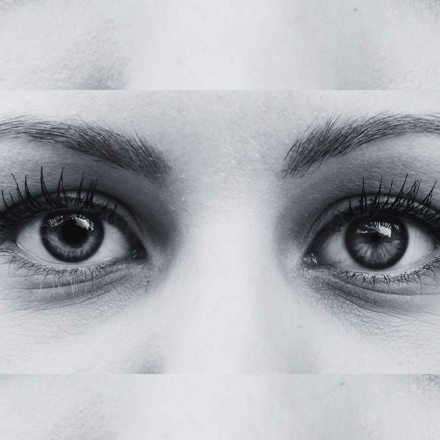
Anisocoria, a condition characterized by unequal pupil sizes, can develop in patients diagnosed with superficial siderosis. The condition is scarce but well documented. The development of anisocoria in superficial siderosis patients can be attributed to the impact of hemosiderin deposition on the oculomotor nerve (cranial nerve III). This nerve is crucial in controlling the muscles that regulate pupil size. When hemosiderin accumulates on the brain’s surface, it can cause progressive damage to the cranial nerves, including the oculomotor nerve. This damage can disrupt the nerve’s function, leading to an imbalance in the control of the pupillary muscles and thus resulting in anisocoria.
Vision Effects
The pupil is the part of the eye that controls the amount of light that enters. It dilates (expands) in low light conditions to allow more light in and constricts (shrinks) in bright light to limit the amount of light that enters the eye. When the pupils are of unequal size (anisocoria), the eye’s ability to adapt to different lighting conditions can be affected.
Here are some potential effects of anisocoria on vision:
- Light Sensitivity (Photophobia): If one pupil is larger and allows more light into the eye, this can increase sensitivity to light, also known as photophobia. This can cause discomfort or pain in bright light.
- Blurred Vision: In some cases, anisocoria can lead to blurred vision. This is more likely if the anisocoria is associated with other eye or neurological conditions.
- Difficulty with Night Vision: If one pupil does not dilate properly in low light, it can make it more difficult to see in dimly lit environments.
- Depth Perception Issues: Although less common, some people may experience issues with depth perception if the anisocoria is significant.
- HeadachesOverview It is challenging to pinpoint the exact cause of su... More and Eye Strain: The constant effort to adapt to different light conditions can lead to headaches and eye strain in some individuals.
It’s important to note that anisocoria often doesn’t cause significant vision problems unless associated with other eye conditions. In many cases, people with anisocoria may not notice any changes in their vision. However, any new, sudden, or worsening symptoms should be evaluated by a healthcare provider or an eye specialist to rule out secondary underlying conditions.
Treatment
The treatment of anisocoria in superficial siderosis primarily focuses on addressing the underlying cause, hemosiderin deposition. The first step is to identify and treat the source of the bleeding to prevent further hemosiderin accumulation. This may involve surgical intervention if a specific source of bleeding, such as a cerebral aneurysm or arteriovenous malformation, can be identified.
Pharmacological treatment may also be used to slow the progression of superficial siderosis and its symptoms. Deferiprone is a drug that has been used to chelate or bind iron in the body, thereby reducing hemosiderin deposition. This treatment has shown promise in slowing the progression of neurological symptoms in some patients with superficial siderosis.
However, it’s important to note that the effectiveness of these treatments can vary, and they may not fully reverse the symptoms of anisocoria. In some cases, additional supportive treatments may be needed to manage the symptoms. These can include the use of corrective lenses or eye drops to manage the effects of anisocoria on vision.
As always, the specific treatment plan for anisocoria in superficial siderosis should be individualized based on the patient’s overall health, the severity of their symptoms, and their personal preferences. It’s crucial for patients to have regular follow-ups with their healthcare provider to monitor their condition and adjust treatment as necessary.
")